SOME COMMON SURGICAL PROCEDURE DETAILS

TPLO (Tibial Plateau Leveling Osteotomy)
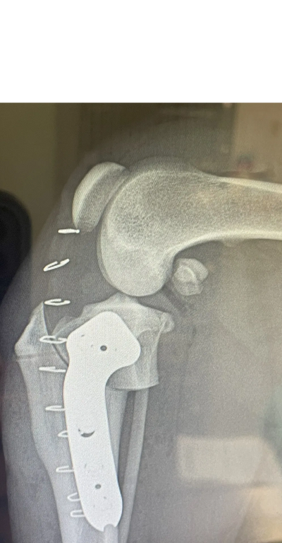
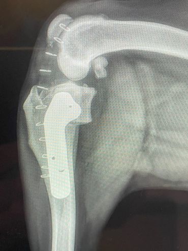
This is a surgical technique used to stabilize the stifle (knee) after a rupture of the Cranial Cruciate Ligament (CCL), which is analogous to the ACL in people. A CCL tear leads to an abnormal back-and-forth (tibial thrust) motion in the stifle which is painful to animals just as it is to people. The instability causes inflammation and the irregular motion on the joint structures causes premature osteoarthritis(OA) to develop at a rapid rate. Once OA has developed it can not be removed and remains for life. To help with OA, Dr. House offers Platelet-Rich Plasma injections(PRP). PRP is injected into the patient's joint to improve cartilage health and also may assist with muscle and tendon healing. To make PRP, your pet's blood is collected and centrifuged to collect their healing factors. These factors are then used in joints and injured areas to encourage healing beyond what the body may typically stimulate. If you would like your pet to receive PRP during surgery, please ask the admitting staff. (See our PRP page for more information.)
Additionally, patients may tear a cartilage in the center of the stifle called the meniscus. There are two menisci in the stifle and typically the medial (inner) one tears which is painful and can cause a characteristic clicking or popping noise when walking. The menisci have a poor central blood supply and generally do not heal well, if at all. During the TPLO surgery, the menisci and joint are normally examined through a larger arthrotomy incision. Dr. House now offers the option of using the NANOSCOPE to perform joint assessment using a tiny hole instead. Using the NANOSCOPE is less painful for your pet and they may walk more comfortably sooner after surgery. If the meniscus is torn however, the joint must be opened to remove the damaged meniscus. If you would like your pet to receive NANOSCOPE arthroscopy, please ask the admitting staff. (See IMAGES BELOW)
If your pet has a torn CCL they will usually be uncomfortable and manifest this as limping. They often prefer to limit their own activity level due to the pain and may be more sedentary. When a CCL tear is diagnosed, usually the best option is to stabilize the stifle surgically for the best outcome. One technique commonly performed for decades has been the Tibial Plateau Leveling Osteotomy (TPLO). The TPLO was designed to eliminate the tibial thrust motion that occurs with a CCL tear and thus return stability to the stifle for return to function. The TPLO procedure is very technical and thus is best performed by experienced surgeons. Initially the TPLO was only taught by the original designer of the procedure, Slocum Enterprises, and luckily Dr. House managed to complete this training program before it ended. To perform a TPLO procedure a bone cut (osteotomy) is made in the shin bone (tibia) near the knee. This cut is curved and allows a change in the angle within the stifle to alter the forces as mentioned above. Once the cut is made and the bone is rotated, a metal plate and screws are placed to hold the bone in its new position while it heals. These implants are usually left in place for life and are only removed if a reaction or infection should develop, which is generally uncommon. Since a cut is made in the bone for this procedure, patients must go through a rest period while the bone heals adequately for normal activity again. It is critical that patients rest to avoid breaking their bone or the implants. The post-operative instructions typically followed are below. If your pet has been diagnosed with a CCL tear, a TPLO may be recommended. This procedure is usually best performed in pets weighing 50 pounds and above. It can be performed in animals under 50 pounds, however, and this can be determined at a surgical consultation. Dr. House performs the TPLO procedure almost daily and would be happy to meet with you and your pet to discuss and perform the procedure accordingly at your local veterinary clinic.
The TPLO Recovery Process
Following the TPLO procedure a strict protocol must be adhered to in order for your pet to heal optimally. A successful surgical procedure is the initial part to a full return to function but without following the recovery plan correctly, your pet could have a poor outcome and could even necessitate future surgical treatment again. YOU MAY DOWNLOAD OUR RECOVERY APP FROM OUR MAIN PAGE TO ASSIST YOU WITH THE RECOVERY PROCESS FOR YOUR PET.
Throughout the recovery period your pet should be restricted indoors when not being monitored. They should avoid going on and off furniture, such as beds and sofas. They should not play with other pets. They should not have the ability to run through your home freely, as a slip could happen or jumping at the door when the bell rings could cause serious injury. Stairs should be avoided. A sling may be used to assist when on stairs, but only minimal stairs should be used. When your pet is not supervised, they should be confined to a room or kennel where they can rest and will not harm themselves.
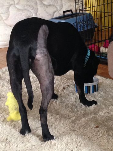
The first two weeks following surgery, your pet must be very limited in exercise on the leash. They may only be taken for short walks outside to relieve themselves on the leash and then must return to restricted quarters indoors. During this timeframe pets should take their pain medications regularly for comfort. The post-operative swelling at the surgical site will improve faster if the medications are used as prescribed, but you may also aid in recovery by cold compressing the incision site. You may cold compress the region for 5-10 minutes every 8-12 hours for 3 days following surgery. Afterwards you may switch to warm compressing for up to 7 more days at the same duration and frequency. You may notice swelling around the hock (ankle) region several days after surgery. This is normal, and a result of gravity, and will resolve on its own or with warm compressing if you wish to speed the process. Finally, during the first 2 weeks of recovery, the external incision site is healing and proper care is paramount to avoid infection and wound opening. An Elizabethan collar (E-collar) is provided and use is highly encouraged. It is critical to avoid letting your pet lick at their incision as only a few licks may instill bacteria into the surgical site and cause a superficial or deep infection. Should a deep infection occur, future surgery to remove the implants will be highly likely. Pets should have the E-collar on always, but should you take it off to feed them, it should be replaced afterwards and they should be watched when it is off. If you see signs at the incision site of progressive reddening, increased swelling or drainage, your should contact your veterinarian promptly. At 10-14 days following surgery your pet will recheck at their regular veterinarian for an incision check and staple removal. If the incision is healing normally, they may have the E-collar removed 2-3 days after staple removal. IN ADDITION TO USING THE E-COLLAR, WE ALSO OFFER THE USE OF THE LICK SLEEVE. The Lick Sleeve covers the surgery leg to discourage licking of the incision. The Lick Sleeve is worn for the full 2 weeks until the incision is healed. It is more comfortable to wear and not in the way. Some pets may CHEW the Lick Sleeve and can damage the incision underneath. It is important to check the incision underneath every day to make sure healing is routine. You may save the Lick Sleeve for future use and can reverse it to use on the opposite limb if needed. If you are interested in the Lick Sleeve for your pet, please ask the admitting staff.
Once the two week recheck has been completed, and your veterinarian is happy with your pet's recovery status, you may now start to perform longer leash walks. Walking on the leash can be done for 5-10 minute periods, 2-3 times daily. This will allow them to burn off energy accumulating from the indoor restriction, and is an important part to faster healing. Walking on the leg will build their muscle back, will increase the joint range of motion and stimulates their bone to heal. When they are not being walked, they should remain restricted inside.
At 8 weeks following surgery, your pet should return to their vet for x-rays. The x-rays are done to assess the bone healing process and to ensure the implants placed have remained unchanged. Your pet's ambulation will also be assessed to ensure healing is occurring as expected. Once this visit is completed, and your pet is doing well, you may then start increasing your leash walks duration and frequency for 4 additional weeks. By the 12 week timepoint after surgery, your pet's bone should be adequately healed to run and play like they used to! Remember that following the recovery process strictly is critical to healing well and that each patient may heal differently. If you ever have concerns about the recovery of your pet, please don't hesitate to ask. We all want your pet to heal well from their surgery and be happy and healthy again!
Nanoscope Stifle Arthroscopy


Lateral Suture Stabilization
This is a surgical technique, which has been around for decades, and is used to stabilize the stifle joint following an ACL tear. The procedure requires an incision on the side of the stifle to place a strong, non-absorbable suture material, which acts as a support so the stifle may heal by scar tissue formation. Although the lateral suture stabilization has been available for a long time, it generally is preferred in smaller patients. Since it requires scar tissue formation for the best healing, patients must strictly rest for 12 weeks afterwards. The major complications are failure of the suture material placed or tearing of the tissues which the suture is supported by, before adequate healing has occurred. The scar tissue healing is generally strong enough to allow good return to function in smaller patients but may not be strong enough in larger (> 50lb) and more active patients. We often select this procedure as it is less involved and thus less costly to perform. The recovery process starts with two weeks of strict rest. The patient may only be walked on their leash for bathroom breaks and then must rest indoors. An E-collar should be used to prevent licking at the incision during this time to prevent infection. Some patients will have a bandage placed to provide additional incision protection and limb support. The bandage may be removed in 2-3 days, especially if it slips, loosens, or becomes wet or soiled. Sometimes the bandage may be left in place until the first recheck. At 10-14 days after surgery, the first recheck is done to check the incision and limb use. The E-collar may come off if the incision is healed. Next the patient may start walking on the leash for 5-10 minute periods, 2-3 times a day for exercise. This will allow them to start building their muscle back, increase the range of motion of the stifle and burn off some energy. At 8 weeks after surgery a final recheck is performed to assess further healing. If healing is going well, the patient may then have increasing leash walks until 12 weeks, at which time they can then resume normal activity. Throughout the 12 week recovery period, it is important patients do not jump on and off furniture, play with other pets or use flights of stairs. If stair use is needed, a sling should be used to provide support. Following the recovery plan strictly is important to prevent injuring the stifle repair which may require a repeat stabilization should injury occur. YOU MAY DOWNLOAD OUR RECOVERY APP FROM OUR MAIN PAGE TO ASSIST YOU WITH THE RECOVERY PROCESS FOR YOUR PET.
Femoral Head and Neck Ostectomy (FHO)
A femoral head and neck ostectomy (FHO) is a procedure on the hip joint. It is used to address many issues including severe hip laxity seen in hip dysplasia, severe arthritis, fractures and dislocations (luxation). The femoral head, the ball, and the associated neck region are both excised with the goal of relieving pain associated with rubbing in the joint. Since the ball is removed, the joint is no longer equivalent to a normal, weight -bearing joint, however the musculature will hold the limb in place. The healing process for the FHO procedure relies on scar tissue formation within the surgical site. While the muscles place the femur in the normal region, the scar tissue forms and stabilizes the area to form a false joint. Full recovery can take up to 6 months, but the exercise restrictions are limited to only 2 weeks. During the first 2 weeks following surgery, strict rest is advised. Strict rest allows for the post-operative swelling to resolve and the skin incision to heal. To assist recovery, passive range of motion may be done on the hip region. Cold compressing may be done for the first 3 days and may then be changed to warm compressing until the recheck appointment. At the recheck appointment 10-14 days post-operative, the incision is assessed and the skin staples are removed, if needed. Exercise restrictions are relaxed and full activity is advised. Activity is important to encourage good limb use, muscle rebuilding and gait training. To attain the best outcome, patients need to use their limb to avoid the forming scar tissue from becoming too rigid and limiting the range of motion. Overall patients having the FHO procedure have a good outcome. They don’t have a normal hip joint, and thus may have an odd step at times, but they are much better off compared to their primary illness. YOU MAY DOWNLOAD OUR RECOVERY APP FROM OUR MAIN PAGE TO ASSIST YOU WITH THE RECOVERY PROCESS FOR YOUR PET.
Patellar Luxation
A patellar luxation, also known as a loose kneecap, is a common condition in dogs and is seen in some cats. It primarily occurs in small breed dogs such as Chihuahuas, Yorkshire Terriers, Shih Tzus and others. The loose kneecap may luxate to the outside (lateral) or inside (medial) locations, relative to the central trochlear groove where it should reside. A medial patellar luxation (MPL) is the most common form. Pain is associated with the kneecap moving in and out of the trochlear groove and lameness will often result. Pets may try to realign their own kneecap by kicking there leg out to the side. If the problem occurs chronically, arthritis will develop and excess stress on the ACL may cause it to tear as well. The condition can be very debilitating and pets may not want to walk, run or play. When the condition causes persistent lameness and pain, surgery is advised for the best outcome. Surgical treatment for the MPL condition involves first deepening the trochlear groove in the joint. The groove is often shallow, contributing the the laxity. Next the tissues that are pulling the kneecap to the inside of the groove are released and then the tissues on the outside of the groove are tightened. Finally the tendon which encompasses the kneecap, the quadriceps tendon, is moved over, to the outside and secured in a new position in the bone below using pins. Realignment can vary in complexity based on the degree of the kneecap laxity. Proper recovery is paramount to a good outcome following surgery. Strict rest is required for 12 weeks while scar tissue forms to stabilize the kneecap after surgery. For the first two weeks after surgery, patients may only be walked on the leash for bathroom breaks. An E-collar is required to prevent licking the incision so an infection does not develop. A recheck is required in 2 weeks to assess the incision and healing. Next the patient may be walked for 10-15 minutes, 2-3 times a day until the second, 8 week recheck occurs. If progress is going well, the patient can have longer walks on their leash and then can return to normal activity at 12 weeks post-op. Throughout recovery jumping on and off furniture, play with other animals and stairs should be avoided to prevent loosening of the tissues and recurrence of the loose kneecap. The pin usually remains in the bone permanently but can rub on the skin, necessitating removal. Overall, the outcome of surgical treatment for patellar luxation is good. For severe cases it may be guarded and advanced corrective bone surgery could be required. YOU MAY DOWNLOAD OUR RECOVERY APP FROM OUR MAIN PAGE TO ASSIST YOU WITH THE RECOVERY PROCESS FOR YOUR PET.
Pet Boarding
Going on vacation? Leave your pets with us! We provide comfortable and safe boarding for your pets while you’re away. Our staff will make sure your pets feel right at home.
Pet Grooming
We offer grooming services to keep your pets looking and feeling their best. Our groomers are skilled in breed-specific cuts and styles. Book your pet’s grooming appointment today.
Pet Adoption
We partner with local animal shelters to help pets find their forever homes. Learn more about our adoption process and meet some of the pets currently available for adoption.
TPLO Images


Videos
A patient walking 2 weeks after TPLO surgery.
A patient walking 5 weeks after TPLO surgery.
A patient walking 8 weeks after TPLO surgery.
This video shows a patient who has had a double (bilateral) TPLO procedure and received the Nocita local pain injection in both stifles (knees). The surgery ended 2 hours prior. Without it they are not as comfortable and ambulatory so soon.
Cookie Policy
This website uses cookies. By continuing to use this site, you accept our use of cookies.